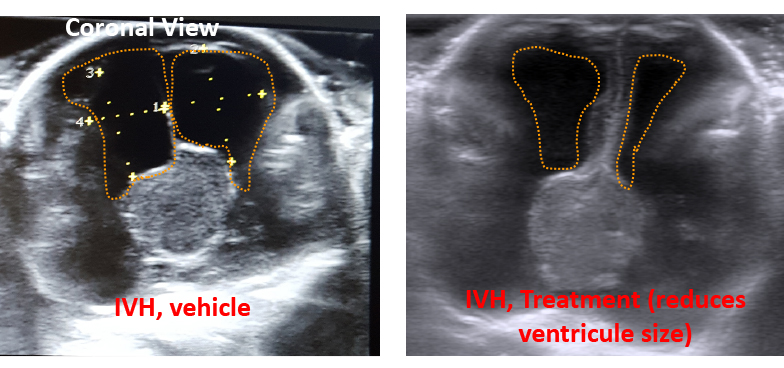
We study the effect of prematurity and IVH on brain development and apply pharmacological and genetic strategies to reverse the cortical injury, white matter damage, and hydrocephalus in the newborns with IVH. To understand the cellular and molecular mechanism of brain injury we employ diverse experimental strategies, including immunolabeling, confocal microscopy, stereological quantification, western blot analyses, RT-qPCR, FACS, bulk and single cell RNA seq, MRI and neurobehavioral studies. For genetic modulation of a target molecule, we employ viral gene delivery system in our rabbit model of IVH. We have recently developed and have characterized a mouse model of IVH, which gives us the ability to use transgenic animals.
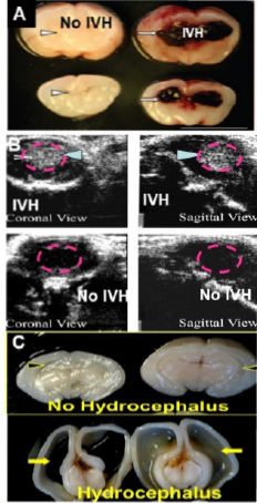
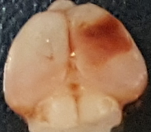
We use E29 rabbit kits (term=32d), delivered by C-section. Rabbits like humans have perinatal growth and larger white matter compared to other rodents. This is a suitable animal model of IVH because kits with moderate-to-severe IVH (like humans): a) exhibit reduced myelination and post-hemorrhagic hydrocephalus b) display an inflammatory response, including elevated cytokines, oxidative stress and microglial infiltration, and c) manifest motor and cognitive deficits. Additionally, we have developed a mouse model of IVH, in which 15 µl of blood is sterotactically injected into the cerebral ventricle of P3 mouse. Blood for cerebral injection is drawn from adult mice.
We have access to a large collection of autopsy materials from preterm infants (>120 brains, postmortem interval <18h). We have IRB approval to collect brain from fetuses and premature infants after their demise. Thus, we are continuing to collect human tissues from Weiler hospital.
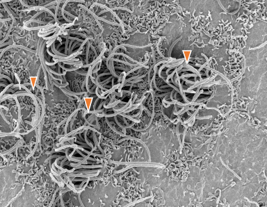
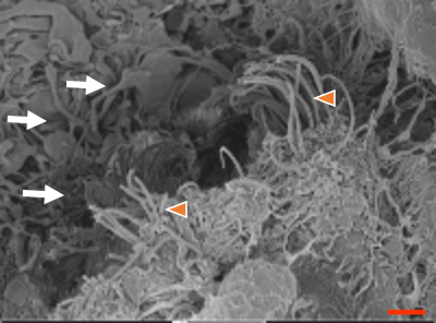
Our present projects are focused on studying plasticity of interneuronal network in premature rabbits and humans and re-constructing the damaged networks in order to accomplish neurological function. In addition, we are studying blood brain barrier, glymphatic system, and ependymal cilia to develop novel therapy for hydrocephalus in the survivors of IVH.